
MOS®, the uniform model for every application
MOSgen™ transforms your samples into MicroOrganoSpheres (MOS): miniaturized, matrix-based 3D models with a selectable number of starting cells, set by simple dilution.
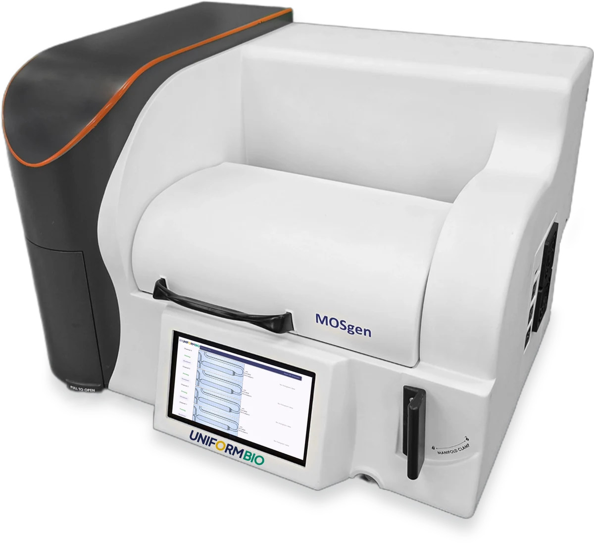
A live run in the chip. Each droplet gels into one MOS.
MOS per run, or 70,000 from a single lane, for 96, 384, and 1536-well plates.
in MOS diameter across a run, in a uniform, matrix-based format.
per run, with no setup, breakdown, or line flushing. Press play, and the MOS come out ready to pipette.
MOS, your single pipetteable format for 3D biology
Your samples, any type, at the cell density you choose. MOS work wherever organoids do, and they pipette like a liquid, so one format serves screening and translational research.
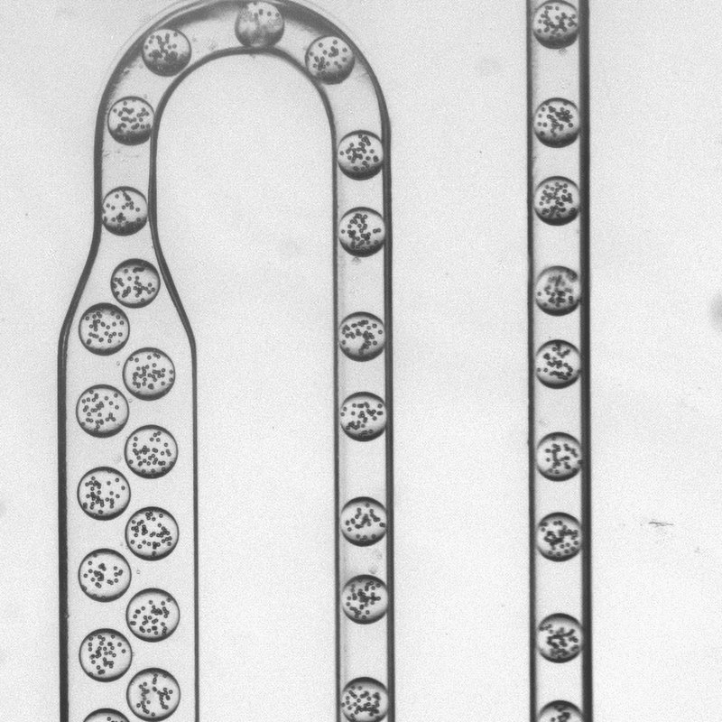
In the chip
Cells in matrix form droplets, up to four lanes at a time.
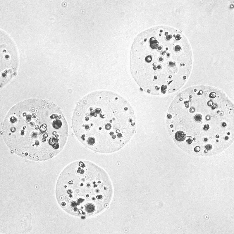
In the well
MOS can dispense into standard multiwell plates.
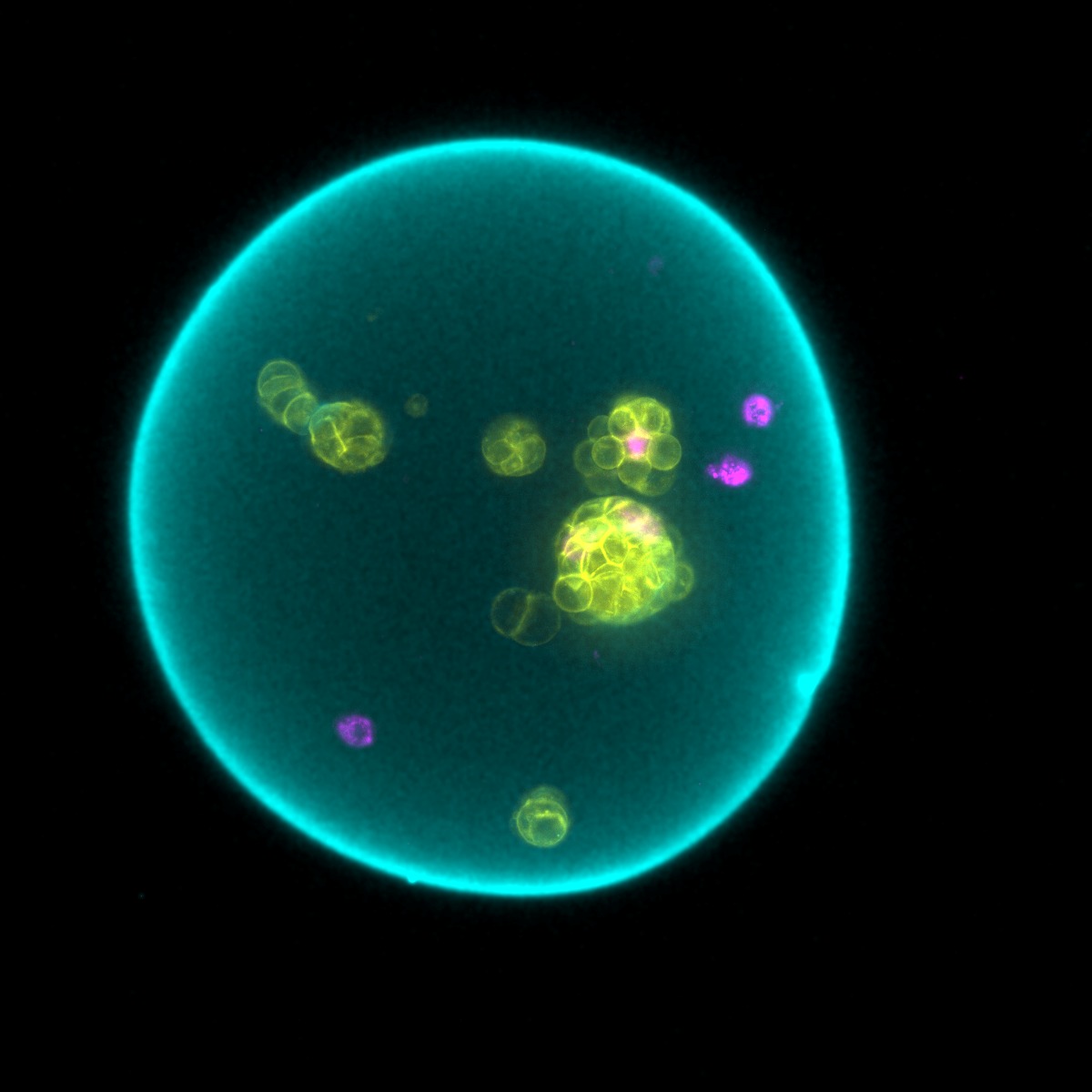
In culture
Cells proliferate and organize within MOS to form 3D models, suitable for every application.
Watch MOS grow in the well
Cells proliferate inside each MOS, and every MOS can be imaged where it sits in the plate.
One well of MOS over 168 hours, imaged every 12 hours. Left: brightfield. Right: fluorescence.
The experiment, from sample to readout
One example workflow. Prepare your samples, and MOSgen makes the MOS. Then use them in any application you choose.
Prep
Cell lines, PDX, PDO, and biopsies, fresh or frozen, as a single-cell suspension.
Load
Cells in matrix and growth media go on the deck.
Scan
Scan the deck and start the run.
Run
Four lanes make up to 280,000 MOS in under 15 minutes.
Dispense
Pipette MOS into 96, 384, or 1536-well plates.
Grow
Cells proliferate and organize inside each MOS.
Treat
Small molecules and chemotherapies, antibodies, T-cell engagers, and cell therapies.
Read
Read each well, by imaging, plate reader, flow cytometry, or sequencing.
Read out on the instruments you already use
MOS are pipetteable. Once they are in the plate, you choose the assay.
Imaging
Brightfield and fluorescence, in the plate.
Plate readers
Plate-reader assays such as CellTiter-Glo 3D.
Flow cytometry
Dissociate MOS back to single cells.
Sequencing
Dissociated MOS go to sequencing as single cells.
ELISA
Measure the proteins MOS secrete into the medium.
Histology
Fix, section, and stain MOS as you would tissue.
Dosing
Add compounds with the liquid handler you run.
Automation
Standard plates, so the rest of the line stays as it is.

3D biology with a 2D workflow
Choose a reason, and we will route your message to the right person.
Thanks, we have it.
We will be in touch to schedule a conversation.
Download the MOSgen flyer (PDF)Inquiry received · routed to the right team